Collie eye anomaly
RoutineOverview
An inherited malformation of the tissue layers at the back of the eye, present from birth and non-progressive in its mild form. It is common in the Rough and Smooth Collie, the Border Collie, the Shetland Sheepdog, and the Australian Shepherd. Most affected dogs are mildly affected and live normally, which is precisely the problem for breeding: a dog can carry and pass on the gene while appearing perfectly healthy. A small proportion develop retinal detachment or haemorrhage and lose sight. Diagnosis is by an ophthalmologist's examination of puppies at around six to eight weeks — the window matters, because the mild changes can become harder to see as the eye matures — and by DNA test. It is not treatable; it is avoidable, by testing before breeding.
What you would notice
- Usually none — most affected dogs see adequately and look entirely normal
- In severe cases, impaired or absent vision, sometimes from puppyhood
- Bumping into things in dim light; reluctance on stairs
Predisposed breeds
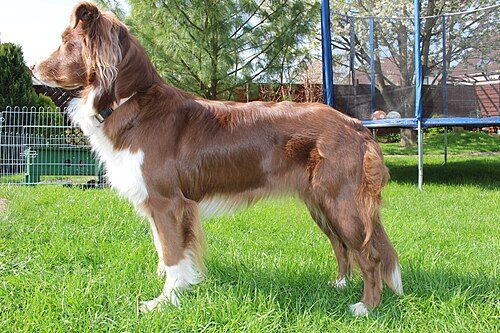
Australian Shepherd
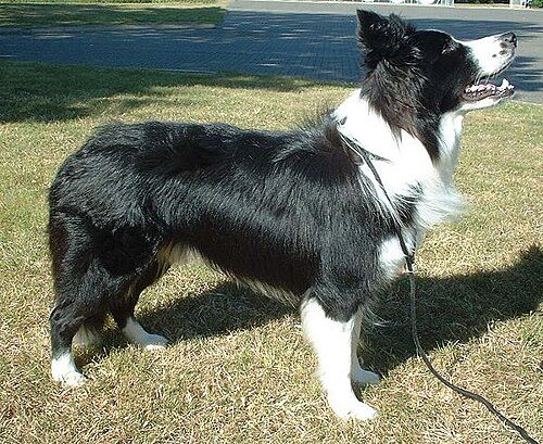
Border Collie
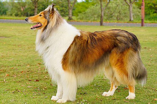
Collie (Rough)
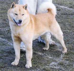
Hokkaido
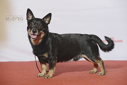
Lancashire Heeler
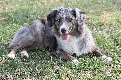
Miniature American Shepherd
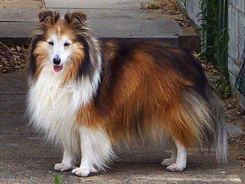
Shetland Sheepdog (Sheltie)
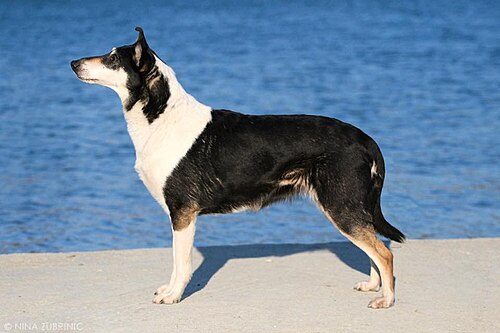
Smooth Collie
Sources
- Merck Veterinary Manual (merckvetmanual.com)
- Orthopedic Foundation for Animals — breed screening databases (ofa.org)